This word describes the process of our joints wearing out (arthritis) due to wear and tear.
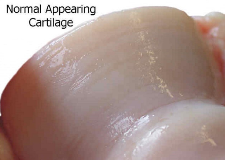
The joint is where one bone meets another. The joints are covered with cartilage on the ends, which form the gliding surface so that the bones (normally) can move one over the other. This cartilage, however, is invisible on the X-ray.


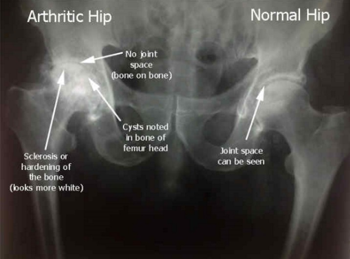
When the cartilage wears out, there are changes that can be seen on X-ray, like in this example showing an arthritic hip and a normal hip.
On the arthritic side, the cartilage is worn away, so it looks like bone touching bone.
The bone gets more stress, and as a result, it gets harder, more calcium, with this process being called sclerosis.
Often, with osteoarthritis, cysts can be seen on the X-ray, as in this picture.
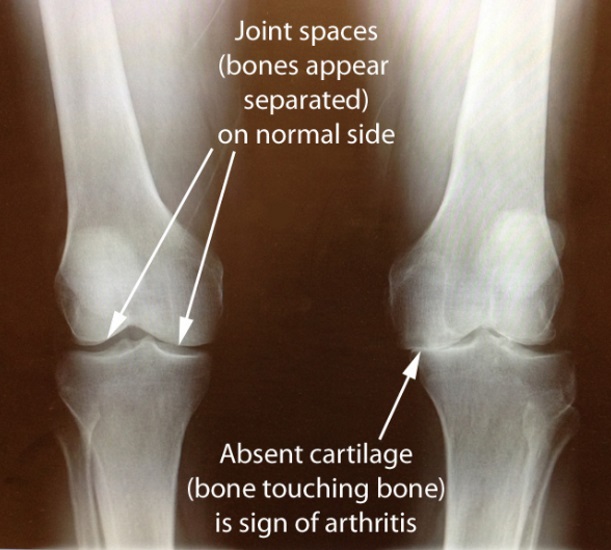
In this standing X-ray of a patient's knees, you can see how on the normal knee that the bones appear separated. The cartilage in between the bones gives the appearance of a space between the bones, since it is invisible to the X-ray beam.
On the side that is worn out, there is no apparent space between the bones, which is an X-ray sign of osteoarthritis.

