Direct Lateral Interbody Fusion
This approach to the spine involves going through the side to reach the disc spaces.
Sometimes it can be difficult to reach the levels at the upper or lower ends of the lumbar spine, but the middle levels can be approached in this manner.
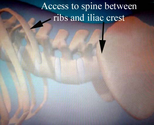
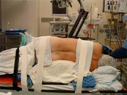
The patient would be positioned on his or her side, and x-ray (fluoroscopy) would be used to help verify the proper level(s) to be treated.
There is a pathway to the side of the spine that goes behind the peritoneum, which is the sac that holds in all of the intestines. This approach goes through the psoas muscle, which is a muscle that flexes (lifts up) the hip joint.
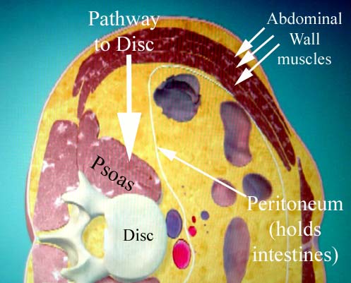
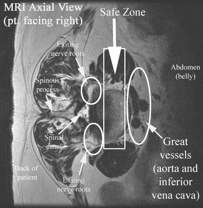
There is a safe zone that is behind the great vessels (aorta and vena cava) and yet in front of the nerve roots exiting the spine.
The psoas muscle is entered with a needle that can be stimulated so that the neuromonitoring technician can tell the surgeon if the pathway is too close to a nerve.
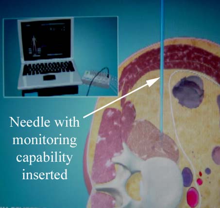
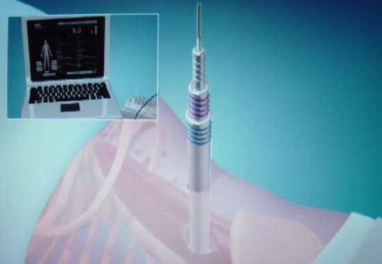
After the safe pathway is identified with the monitored needle, dilating tubes are placed over the needle to provide a path to the disc space.
The retractors are put in place, and after a thin bit of muscle is cleared away, the disc can be seen. the side of the rim (annulus) of the disc can be opened to start clearing out the disc material
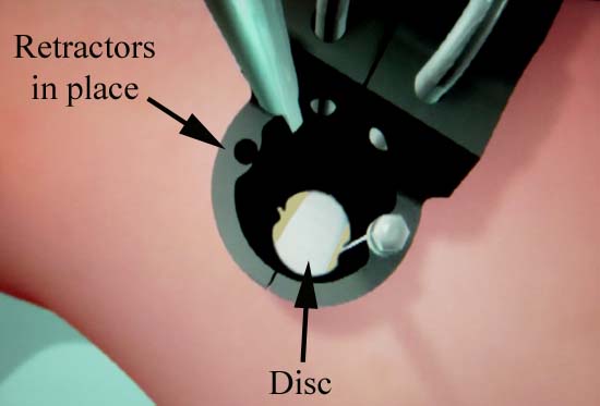
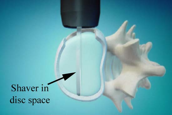
Much of the disc material can be removed with a shaver.
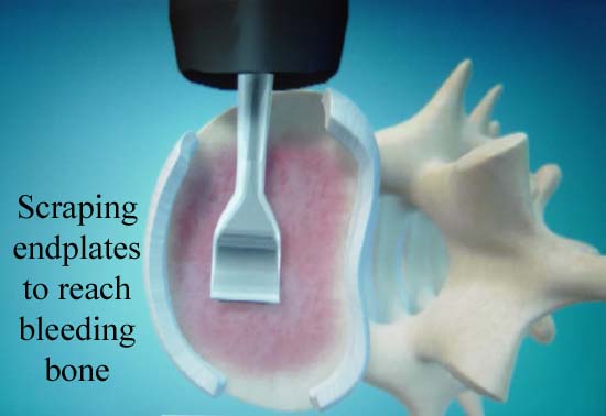
The endplates of the vertebral bodies are scraped to reach bleeding bone, removing the cartilage, but not penetrating the bony endplate. This action creates a surface for fusion, but if disc material is left in place, fusion will not likely occur.
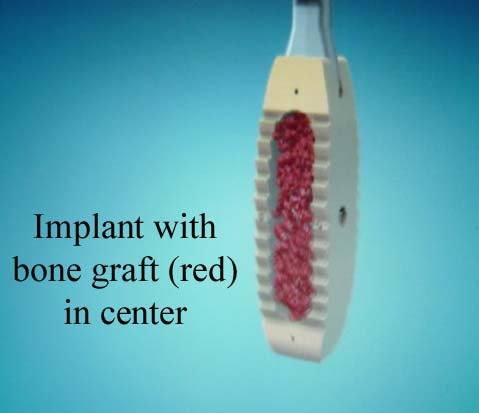
The implant to go in the intervertebral disc space is made of a special type of plastic, but has a hole in the middle to contain the bone graft.
This schematic shows the implant inserted.
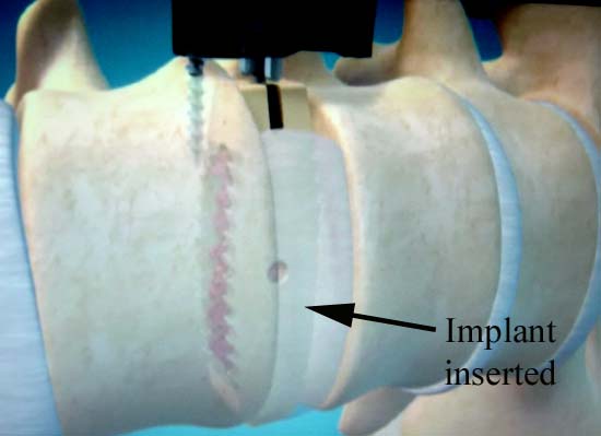
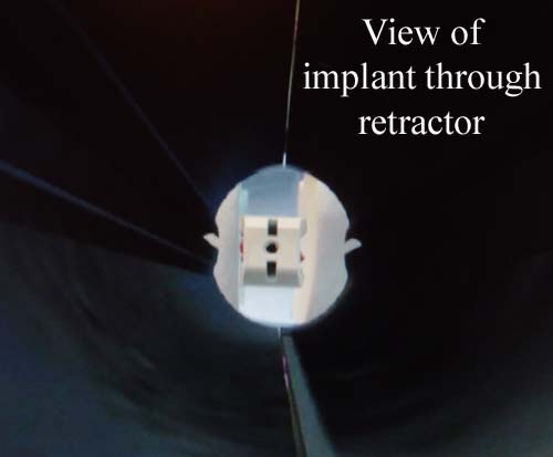
Here is what the side of the implant would look like, viewed through the retractors.
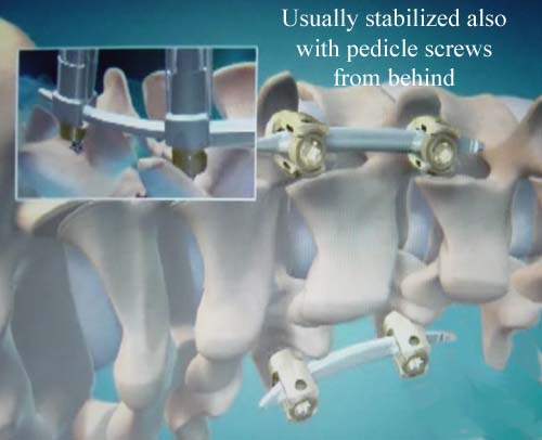
Usually the implant by itself is not stable enough to heal on its own. In most cases, screws are placed in the spine from behind (pedicle screws) or a spinous process clamp to provide additional stability.
All these approaches can be done with a minimally invasive fashion.
An animation is available at the Medtronic site.
