Ankle Arthritis (Osteoarthritis)
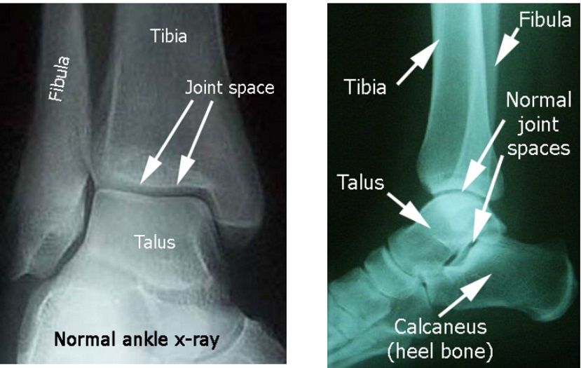
The ankle is the joint where the bones of the leg, the tibia and the fibula, meet the talus, which is the bone above the heel bone. Normally, the surfaces of our joints are covered with cartilage, which is invisible to the x-ray beam, so there is the appearance on x-ray of a space between the bones. There is not actually a space there, but it looks that way since the cartilage can’t be seen. Here are some normal appearing ankle xrays:
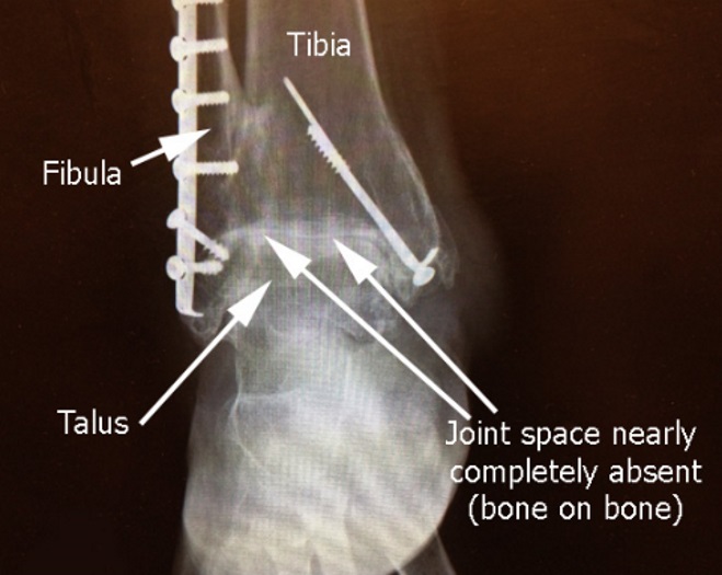
Sometimes, the cartilage of the ankle can wear away. This condition can occur due to multiple reasons, but the most common are trauma and inflammatory problems, like rheumatoid arthritis. Lack of joint space can be seen in the x-ray to the left.
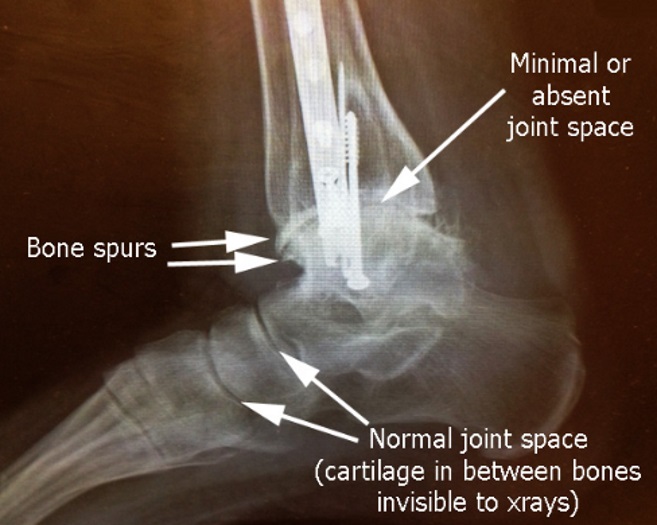
In the lateral (side view) of the same patient, there are bone spurs on the front of the ankle and no joint space between the talus and the tibia.
This loss of cartilage means that instead of the joint moving smoothly and painlessly, the surface becomes rough, with the bone ends beginning to grind together. The result is a decreased range of motion, with more pain and disability.
Treatment Options
The choices for treatment:
-Do nothing
-Medications
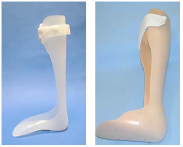
-Mechanical support (ankle foot orthosis) pictured left
-Surgery
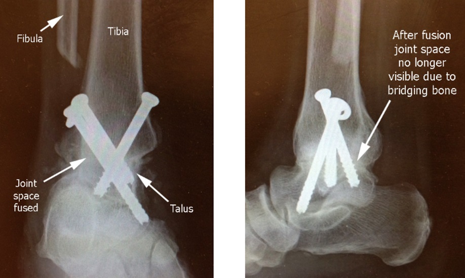
Surgery: usually the option is called Ankle Fusion, pictured here, where the arthritic bone surfaces are removed, and the bones are held together with screws. The biologic situation is created where the body reacts as if it were a fracture and tries to heal the two bones into one bone. The fusion procedure is like gluing the two bones together with bone.
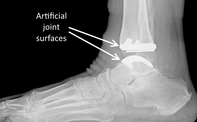
Another option might be ankle joint replacement, or artificial ankle. For this joint replacement procedure, however, there is a lot of stress concentrated on a small surface area, and the ideal patients would be those with low activity demands.
For patients interested in that procedure, a referral can be facilitated (as I don't do this type of surgery).
