Cervical (neck)
Spondylotic (spine wearing out)
Myelopathy (spinal cord dysfunction)

This term describes a condition in the neck (cervical region) where the spine gets so degenerated (spondylosis, or the adjective form of the word is spondylotic) that there is impingement or squeezing of the spinal cord causing spinal cord dysfunction (myelopathy).
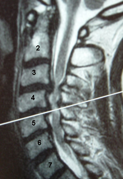
These views, through an unaffected area, C2-3, show how the spinal cord has plenty of room around it with the spinal fluid, which shows up white in this MRI sequence.
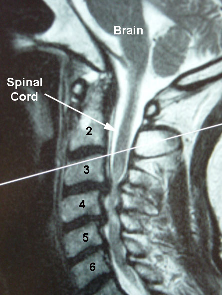
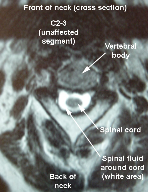
However, at the C3-4 area, which is more severely involved, shows no room for spinal fluid around the cord, as the spinal cord is compressed by the disc bulging and other degenerative changes. This patient at the time of presentation had severe spinal cord dysfunction with balance issues and abnormal reflexes.
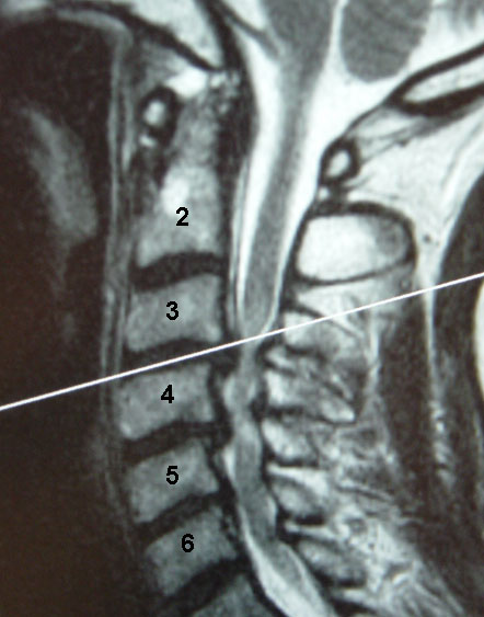
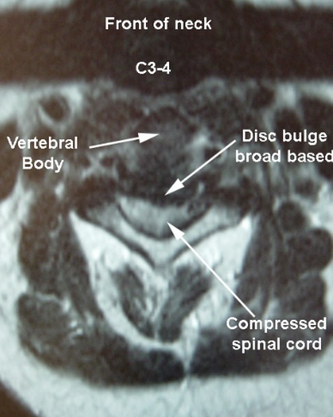
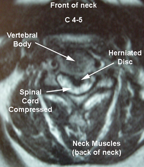
Similar findings are seen at the C4-5 level as illustrated below, with a large herniated disc pushing right into the middle of the spinal cord. While the spinal cord normally on cross section has an oval shape, here the compression make the cord have the shape of a kidney bean (not a good thing!)
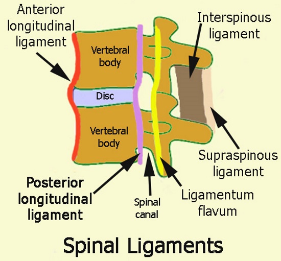
The Posterior Longitudinal Ligament (PLL) lines the back side of the vertebral body, and is at the front side of the spinal canal. This ligament rests right against the front part of the dura, which is the sac that contains the spinal fluid and the spinal cord.
In the diagram to the right, the PLL is illustrated by the purple line.
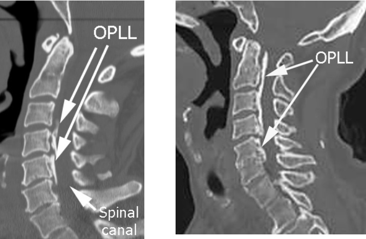
Another condition that could cause spinal cord compression is OPLL: Ossification (calcifying, or turning to bone) of Posterior Longitudinal Ligament
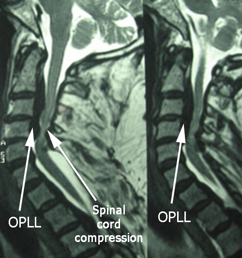
In these cross section views on MRI studies, the thick black line behind the vertebral bodies, which in this case is composed of calcified tissue, looks black on the MRI scan. The presence of the OPLL here contributes to the compression on the spinal cord. At the areas of compression, there is no white fluid around the spinal cord as there is above and below.
In the images to the right, which are from CT scans, the ligament tissue which has ossified (turned to bone) shows up white.
Clinical Significance of Myelopathy
The spinal cord itself, which is part of the central nervous system, is not a structure which when compressed causes a patient to experience pain. Rather, there may be more subtle symptoms with spinal cord compression: balance issues, problems with fine motor movements (buttoning clothes). Also, there are certain findings the doctor will note on exam, like abnormal reflexes and coordination deficits.
Myelopathy tends to be progressive and in most cases, surgical treatment is recommended.
The clinical significance of OPLL regards planning a potential surgical approach. While in most cases, the spinal cord can be decompressed most directly from the front approach (as illustrated here), in cases with OPLL, usually the decompression has to be done from the backside, or what is called a posterior approach, such as with laminoplasty or laminectomy. The formation of bone in the PLL that occurs in OPLL often causes the bone to become adherent to the dura (the sac containing the spinal fluid), so that operating from the front has a higher chance of having a spinal fluid leak.
