Pain in the neck can be from any of a variety of structures, if injured or worn out: muscles, fascia (the tissue covering the muscles), ligaments, discs, bones, joints, and nerves.
When the pain is from torn muscles or fascia (the sock covering the muscles), there is usually tenderness to touch in the involved areas. When the pain is from a disc or ligament or joint problem, which are deeper structures, it is usually difficult to reproduce the pain by touching the back of the neck.
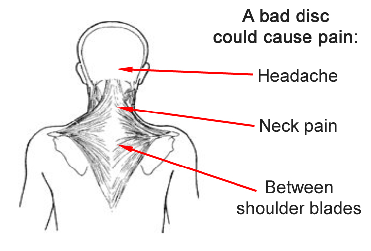
When pain is from cervical degeneration, patients can have pain in the neck, between the shoulder blades, and/or headache.
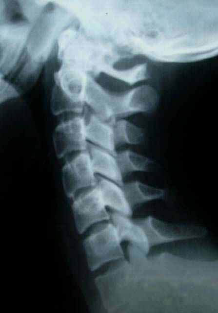
To appreciate the types of abnormalities that can be seen on xrays, look first at this cervical spine xray that would be considered normal.
The disc spaces, the cushions between the bones, are all about the same thickness in this x-ray to the right.
Note also the curve of the cervical spine: that there is a slight bow forward of the neck bones, with a curve that is concave (the hollow side) towards the back.
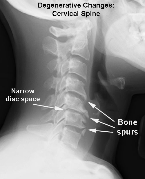
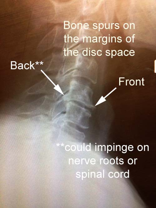
The x-ray below shows degenerative changes with bone spur formation and narrowing of the disc space (think of a water bed with the water drained out).
Bone spurs are easy to see on the front of the disc space, but in this example, they can also be seen on the back side of the disc space, where they can cause compression of the nervous structures including the spinal cord and/or the nerve roots.
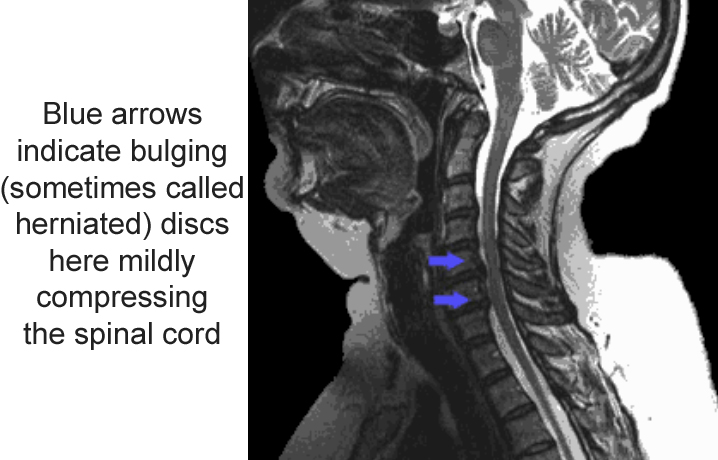
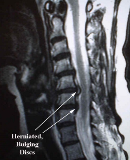
In this MRI to the right, looking at the cervical spine from a sagittal (side) view of a different patient, the arrows indicate bulging or herniated discs that in this case are touching the spinal cord.
In this case, without the MRI, we could not have seen the herniated disc at the C3-4 level. Note that the disc height is not significantly decreased at the level without the disc bulge contacting the spinal cord.
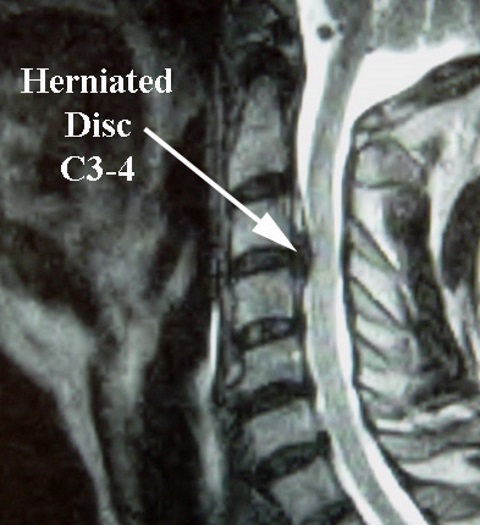
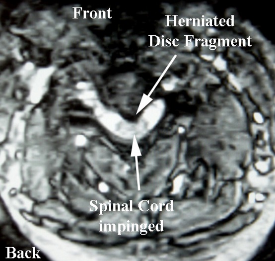
On the cross sectional view, the C3-4 herniated disc pictured on the side view above can be seen indenting and impinging upon the spinal cord.
Since there is not much extra room around the nerves and spinal cord, it would make sense that a small mass (disc material) next to the nerve structures can cause significant symptoms.
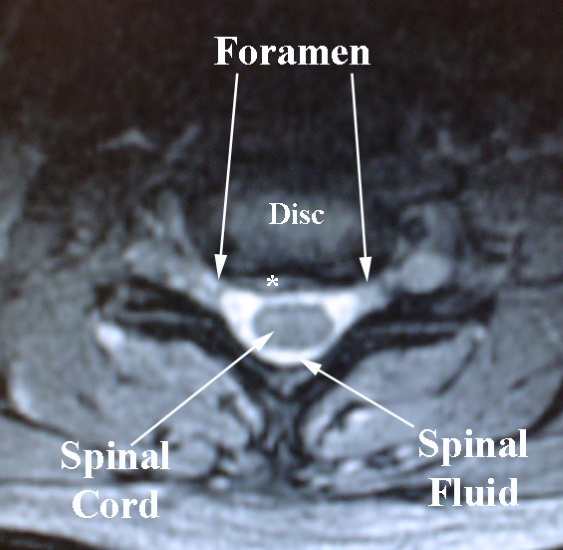
Another example: here is a section through a segment without symptoms.
There is a small disc bulge marked by an asterisk, but this bulge is not likely causing problems, as there is no pressure on the spinal cord or an exiting nerve root.
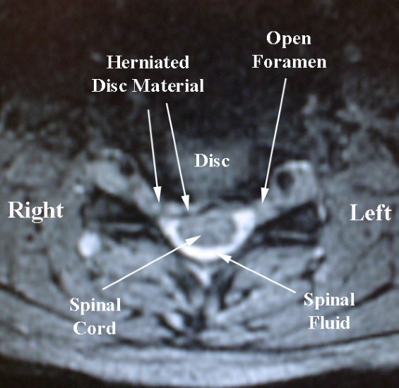
However, at another spinal level on the same patient, there is a large herniation at the level pictured here.
This extruded disc material almost blocks the path for the exiting nerve root on the right side, which correlated with the patient's right arm pain and numbness.
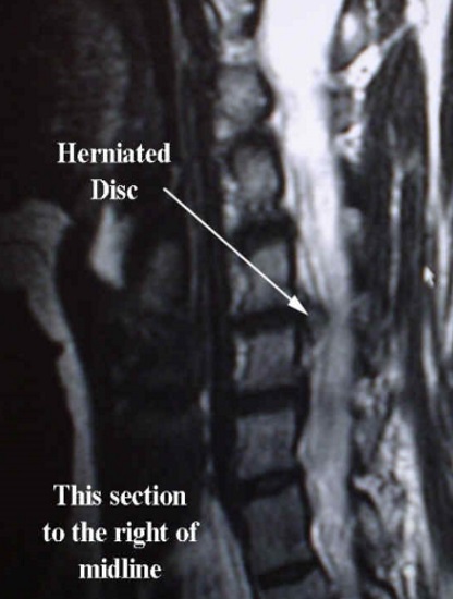
In the MRI sagittal image (looking from side view, slicing left and right in half) below left, the herniated disc (disc out of place) can be seen.
In the MRI sagittal image below right, with this slice showing structures on the same side of the disc bulge noted above (right side), the bulging disc can be easily seen, and correlates with the patient having right arm pain.
