Spinal Stenosis (Narrowing of the Spinal Canal)
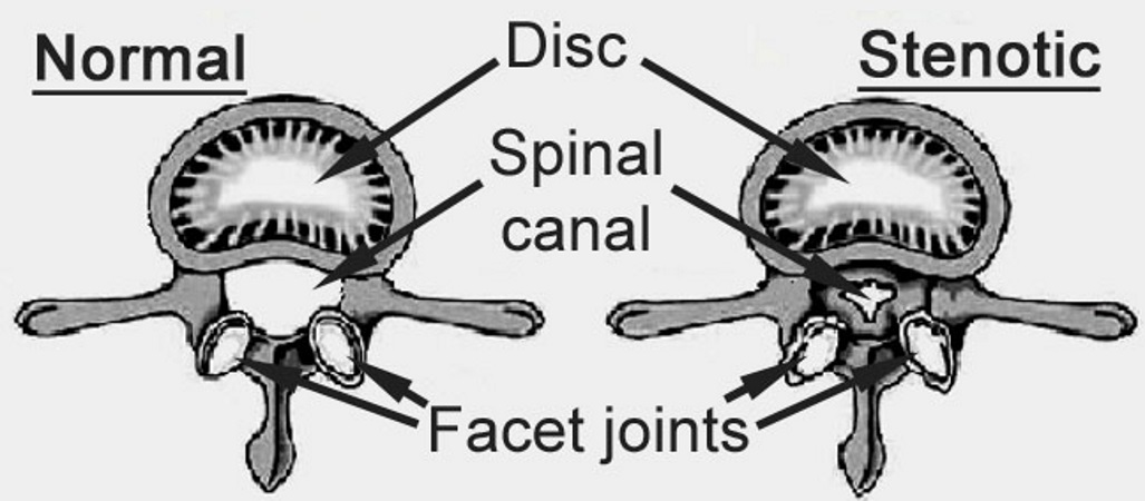
Spinal stenosis is a condition, where due to some type of process, either degenerative (wear and tear), for reasons related to either the discs or facet joints, or because of a change of position of the spine bones, the space for the nerves becomes more narrow. In the illustration below, the area marked “spinal canal” is where the spinal nerves are located.
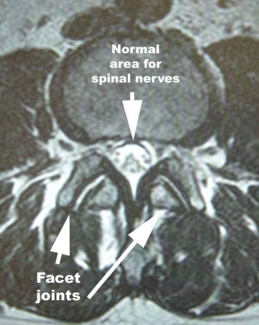
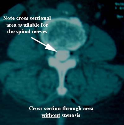
MRI cross section without stenosis or narrowing of the spinal canal.
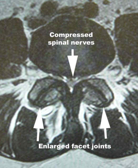
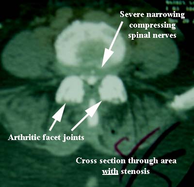
MRI cross section with stenosis or narrowing of the spinal canal.
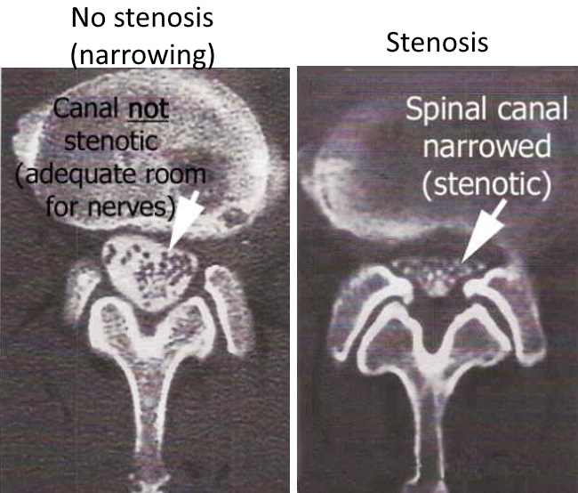
The images here are from a CT myelogram, which is a test where radiographic dye (the white material in the spinal canal highlighting the nerve roots) is injected into the area occupied by the nerves, to help show how much room there is for the spinal nerves.
The vertebral bodies are in the upper part of these cross sectional images, which show no significant narrowing in the image with black letters, but does show narrowing/stenosis in the image with text in white.
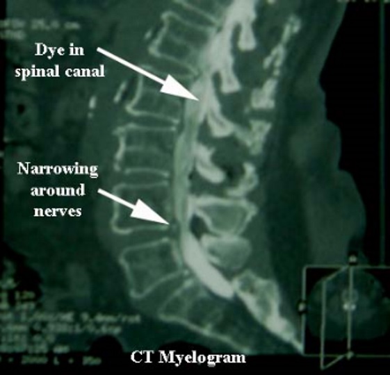
What often will occur in degenerative conditions is that due to the combination of the disc settling (think of the disc as a jelly donut, having the jelly dry out), the arthritic processes of the facet joints, and the subsequent infolding of the ligaments between the bones, the area for the nerves becomes too narrow.
In the image to the right, a sagittal or side view, shows the spinal canal behind the vertebral bodies, and narrowing can be seen here at the L4-5 level.
For more information on facet arthrosis, click here.
Cross sectional views are seen below.
The center image shows no stenosis/narrowing.
The image on the right shows spinal canal narrowing/stenosis.
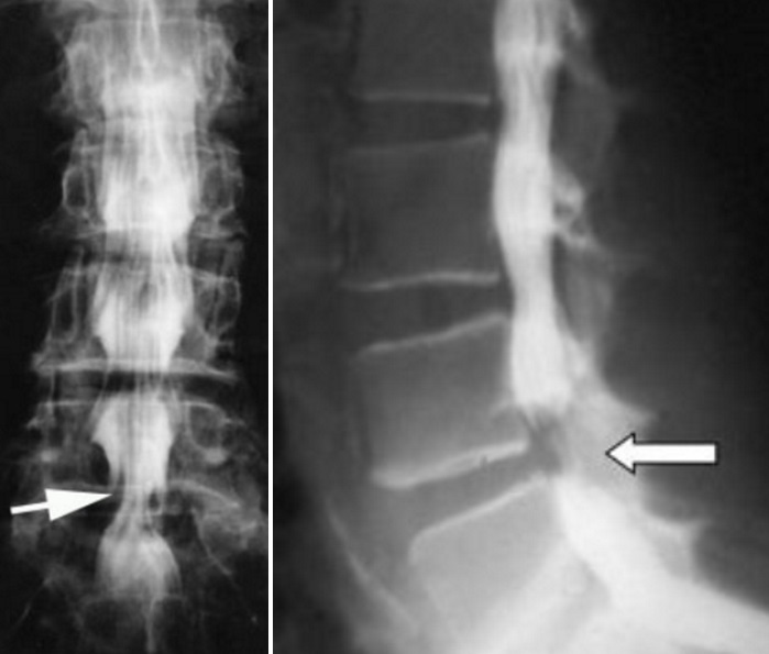
In these myelograms, which are studies where contrast dye is injected to mix with the spinal fluid, deep to the dural layer (like for a spinal anesthetic), the narrowing can be seen lower in the spine as indicated by the white arrows. Without this injection of contrast, the narrowing around the nerves can be difficult to see.
Symptoms:
The effect on the nerves can best be understood by thinking of the analogy of a belt around your waist: if that belt started to tighten, the soft tissue underneath (your waist in this example) is compressed.
Eventually, a person develops symptoms that frequently include heaviness in the legs, fatigue, leg pain, numbness, and weakness. Frequently, the symptoms are made worse by walking and standing, and better by leaning forward (like on a grocery cart when shopping, or sitting. The upright posture causes a small amount of increased compression on the nerves, and forward flexion gives a little bit of relief of the spinal nerve compression.
These changes usually occur in patients over 50, and symptoms can develop very slowly, often over years.
While non-operative treatments such as medications, physical therapy, and injections can help some, surgery is frequently needed to relieve the pressure on the nerves.
When non-operative treatments fail to help, often the patient will choose a surgical option, such as laminectomy, which is usually an open procedure, or with a minimally invasive approach, a microdecompression can be done in many cases.
