Lumbar Fusion Case Examples
Patient is 52 yr. old male with chronic severe low back pain that has failed to respond to conservative (non-operative treatment), including medications, therapy, and injections.
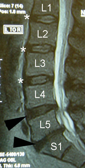
On the lateral (side) view of the pre-op MRI, note the normal appearing discs between L1 and L4, marked with white asterisk. The discs between L4 and S1, marked by the dark arrowhead, are not normal appearing. While the disc at L5-S1 is clearly flattened, the disc at L4-5 has a bulge at the back and does not have the same white signal as the normal discs above.
The decision was made to include the L4-5 disc in the fusion since, after fusing L5-S1, the stress would be concentrated on the next level, which in this case, already shows signs of wear and tear.
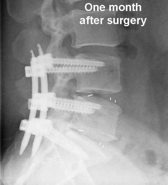
One month after surgery, the interbody spacers, which are invisible to xray except for the metal markers in them, can be seen in the interspaces between the vertebral bodies.
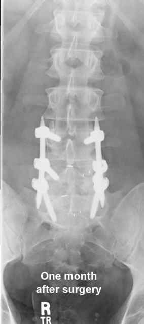
The patient’s back pain was significantly better by about 6 weeks.
Seven months after surgery, the patient is feeling much better. On the lateral (side) view, some calcification (whiteness) can be seen in the interspaces. This finding, however, is subtle.
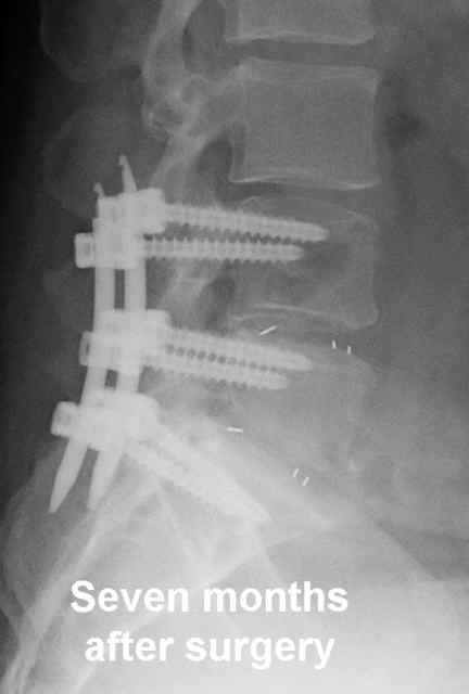
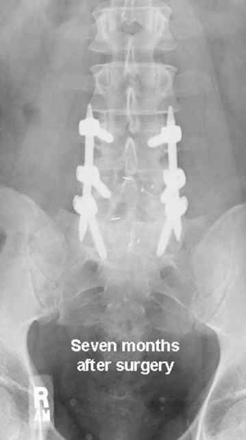
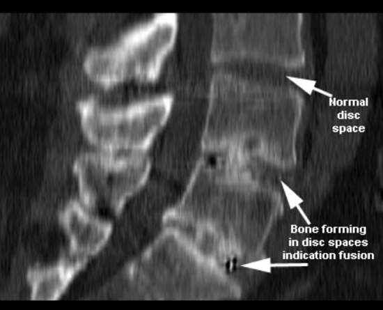
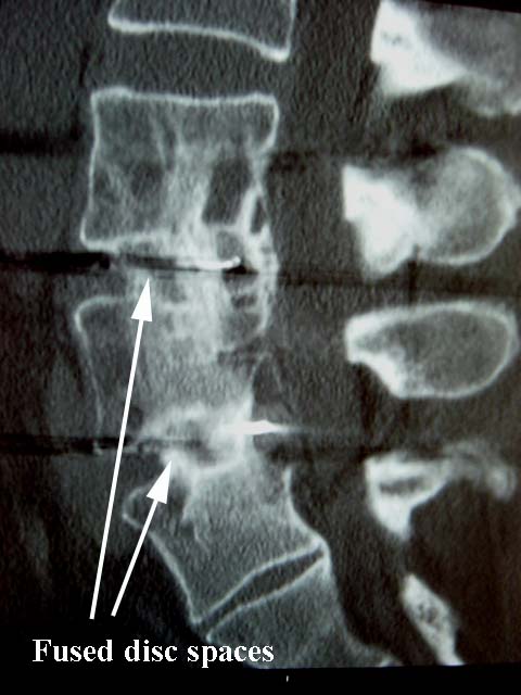
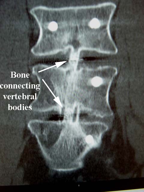
By checking the patient with a CT scan, however, the bony healing between the vertebral bodies can be demonstrated on the reconstructed views.
The patient was released to his regular duty at 8 months post op. However, since he has two less discs in his spine (four discs doing the job of six), he is still advised to avoid frequent bending, lifting, and twisting.
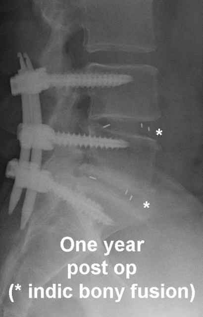
On the lateral X-ray at one year post op, more calcification can be seen between the vertebral bodies as indicated by the asterisk.
The patient continues to do well.
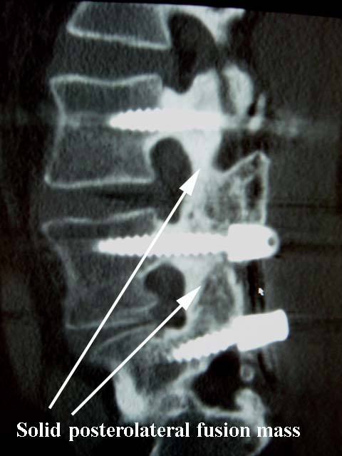
Here’s another example of a patient who at one year after a two level fusion had a solid bony connection.
However, here is an example of a case where the fusion did not occur (nonunion). The bone is growing from above and below within the spacer, but it never connected. This example is shown since, even with preparation of the endplate and removal of disc material, sometimes fusion does not occur. The patient didn't do anything wrong. Sometimes these outcomes occur.
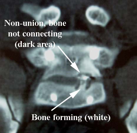
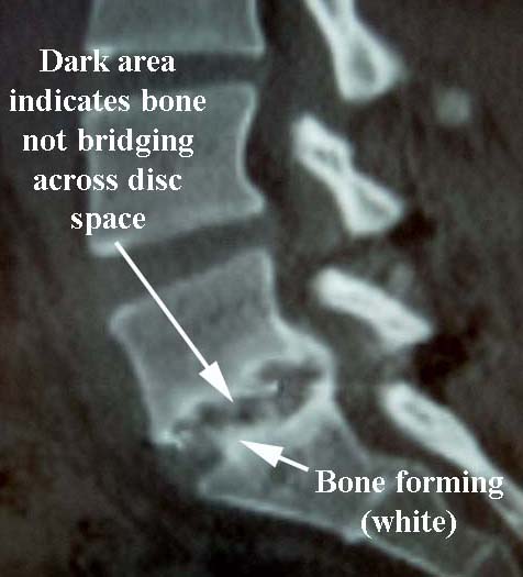
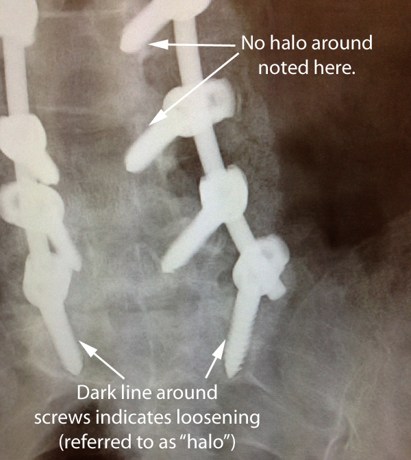
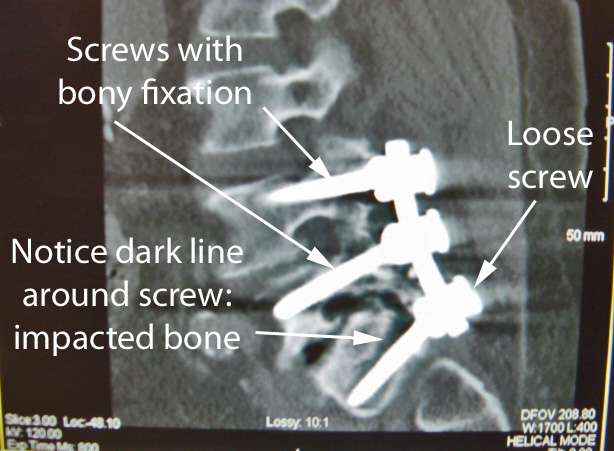
On those occasions where a fusion does not fuse (nonunion), the screws will eventually become loose. In the X-ray below, you can see that the two lowest screws have a dark line around them that is referred to as a “halo.” Although the screw will have a good bite (or purchase) on the bone when it is initially placed, when it gets loose, it will to a small degree compress the bone around the screw. This absence of bone around the screw has the appearance of a dark line around the screw. In the x-ray below, the screws higher in the spine, which are not loose, do not have the same halo.
This image to the right from a CT scan, taken from a different patient, shows the x-ray appearance of screws when they become loose. This loosening is demonstrated by the dark line around the screws (referred to as a “halo”), which indicate that the screw threads have no purchase or bite into the bone.
Keep in mind that the purpose of the screws is to hold the bones in place until the healing (fusion) occurs, and that even in the best circumstances, the fusion rate in the lumbar region in large groups of patients is somewhere between 80-90%.
However, not every patient with a CT scan like this requires revision surgery: each case is considered individually.
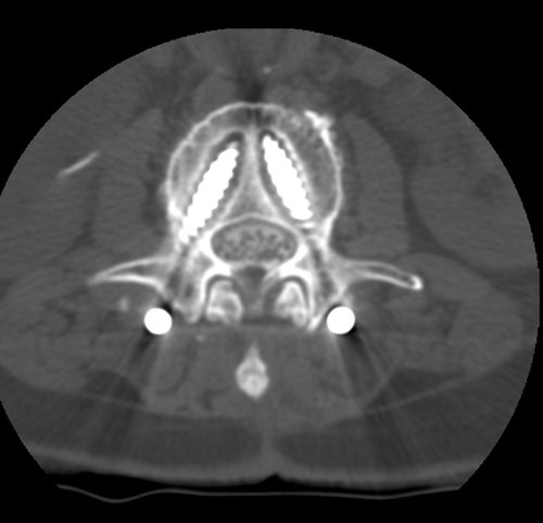
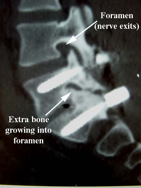
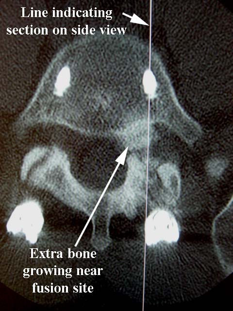
Here is a case where extra bone grew near and in the foramen, which is the opening through which the nerve root exits the spinal column.
In this case, although the patient might be expected to have pain along the area supplied by the involved nerve root, the patient had no symptoms. This case also illustrated why the focus is on treating the patient, not the x-ray.
