Foraminal Stenosis (Narrowing)
The foramen is the hole or opening through which each nerve leaves the spinal canal.
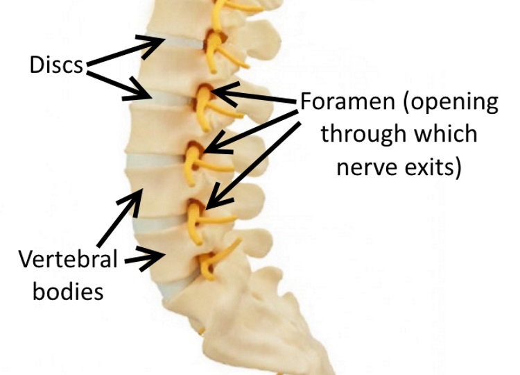
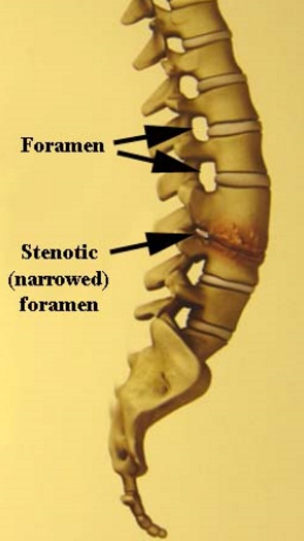
Stenosis is a word meaning narrowing.
Foraminal stenosis is a condition illustrated here where, due either to bulging of the disc in the front of this opening, or from arthritic change of the facet joints from the back side of the opening, the exiting pathway for the nerve gets more narrow, squeezing the nerve.
Symptoms, which usually include pain down the leg for low back conditions, or down the arm for cervical (neck) conditions, are usually worse when standing upright, as the opening usually gets smaller in this position. By leaning forward, which flexes the spine, the opening gets a little bigger and sometimes patients can get some relief from this position change.
This MRI view of the lumbar spine is a side view, but instead of looking at the cut right down the middle, this cut is off to one side which helps to visualize the foramen at each level.
In this MRI study to the right, normal levels (with fat as a cushion around the exiting nerve root) can be seen above and below a level where the exiting nerve root is compressed (no white fat around the root, since it is compressed by a bulging disc).
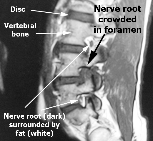
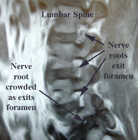
In this MRI from another patient, the top three foramen are normal in appearance with white fat surrounding the exiting nerve roots.
At the lowest level, however, which is stenotic (narrowed), there is no fat surrounding the nerve.
If the narrowing around the nerve is significant, no positional change will give relief.
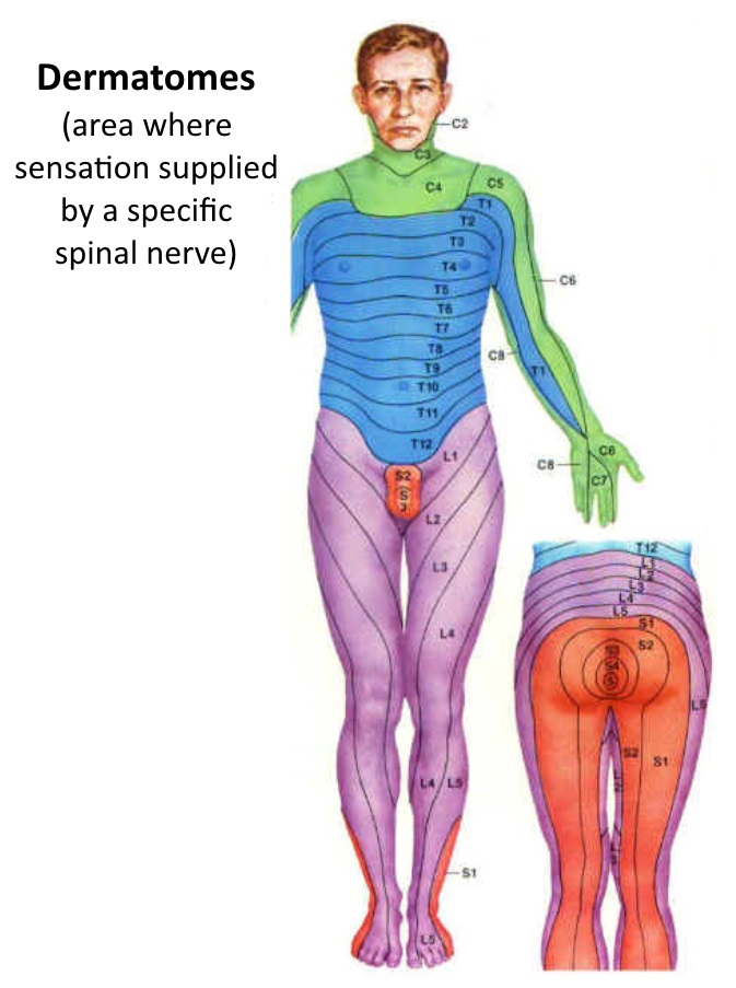
The discomfort perceived from compression of the involved nerve root is usually experienced in the area of the body supplied by that particular nerve.
Just like when you hit your funny bone in your elbow, and that tingling feeling goes into your little finger, each nerve that exits the spine has its own distribution, or area to which it gives sensation.
For any individual nerve root, the area of the body where, if that nerve were compressed, the patient would feel some tingling or pain, is called the dermatome. The dermatomal distribution or coverage of the various nerve roots is noted in these diagrams.
Very often, since medications or cortisone injections don’t make the opening bigger, surgical treatment in the form of foraminotomy is required.
