Lumbar facet arthrosis
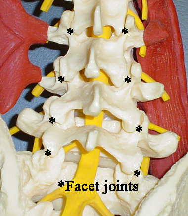
The facet joints are in the posterior (back side) aspect of the spine, and are to the right and the left from the midline. These joints, which have a sliding motion to allow the spine to flex forward and lean backward, have surfaces covered by cartilage, like on the end of a chicken bone.
These joints are indicated in the picture on the right by the asterisk (*) on the photograph of the spine model.
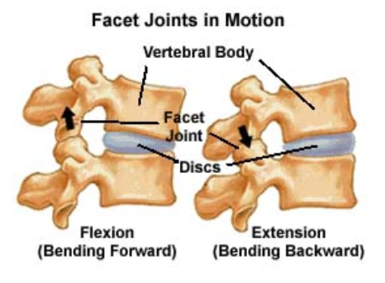
When a patient bends forward and backward, the disc remains the center of motion, but the back part of the spine has to get either longer (to bend forward) or shorter (to bend backward). These motions are accommodated by the facet joints which normally have smooth surfaces.
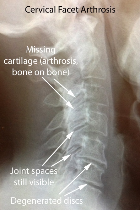
Part of what occurs with arthritic change is that the cartilage wears away, and the surfaces become rough and irregular instead of smooth and low friction.
These joints, just like a knee or hip, can become arthritic. The cartilage can wear away, and the result can be bone rubbing on bone.
When this process occurs, the body reacts by forming more bone which on x-ray would look like a cloud or a cotton ball forming over the involved joints, which would look more white on x-ray.
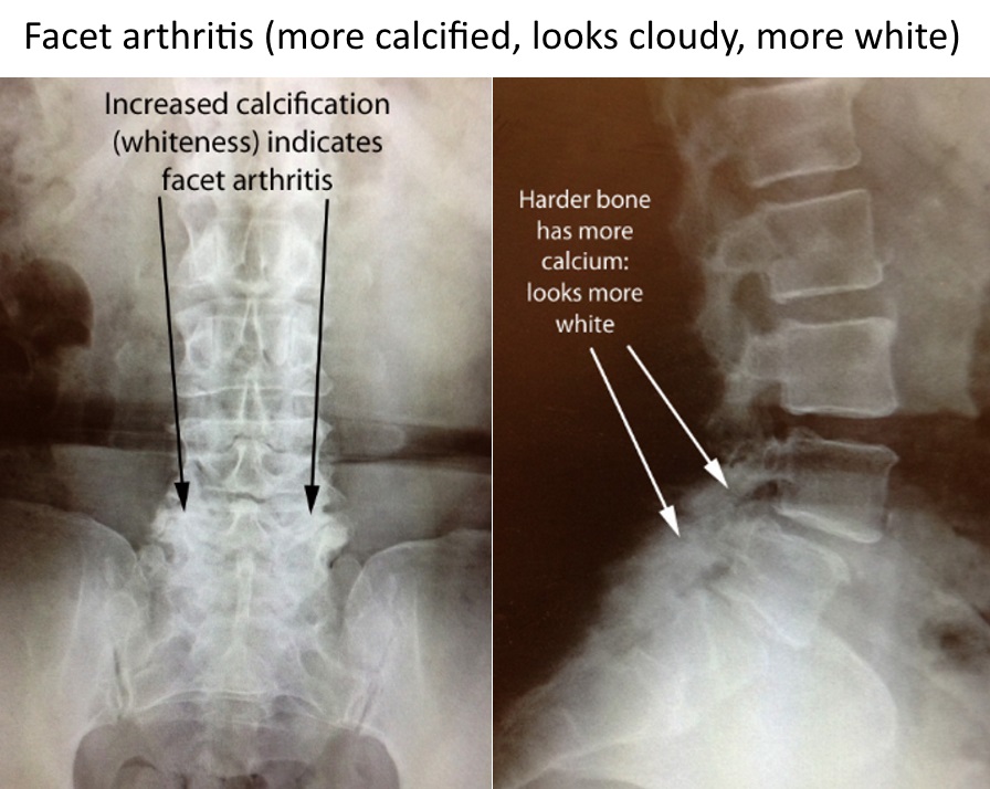
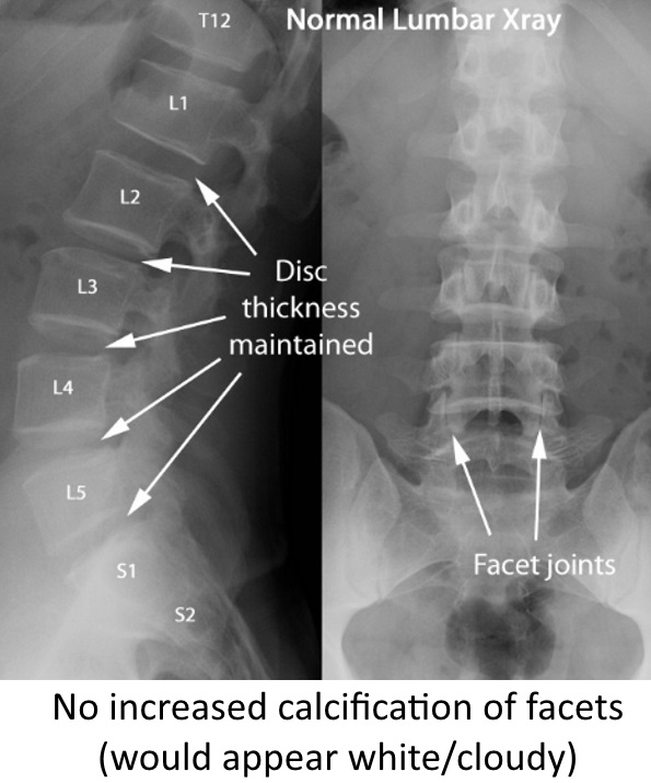
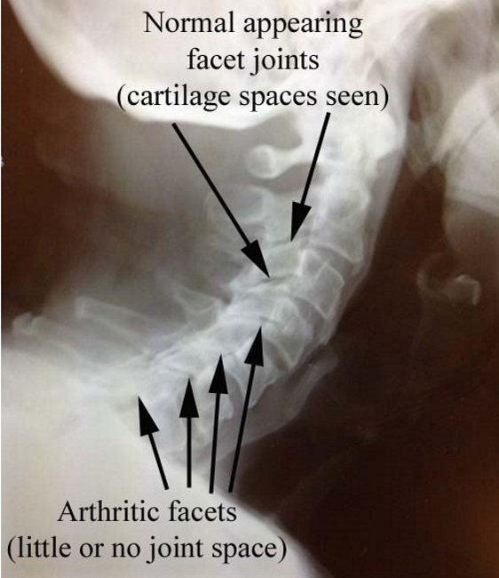
Compare the arthritic x-rays above with the normal x-rays here, without the calcification of the facets.
Cross sectional imaging
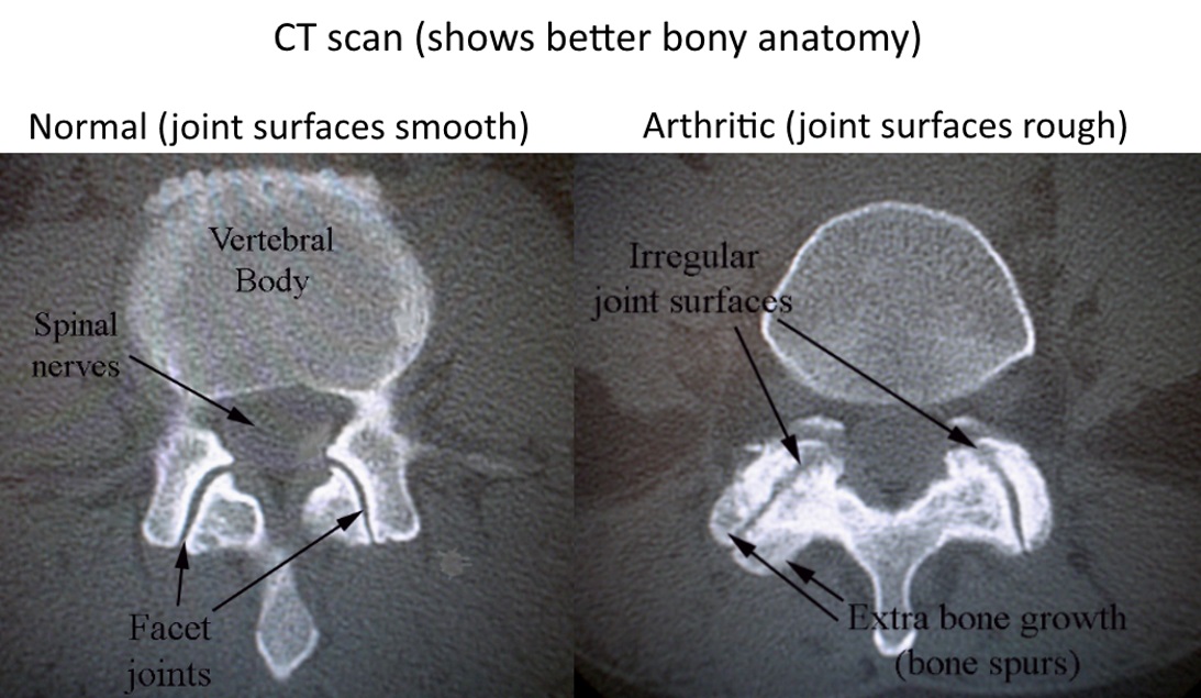
A better way to look at these joints is with a three dimensional study, like an MRI or CT scan, that can make images in different planes/directions.
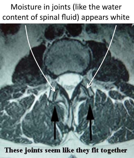
In this cross sectional view which shows normal facet joints, notice that it appears that the two bones that form the facet joint fit nicely together.
There is a thin line of normal appearing joint fluid between the bones of the facet joints. Think of this thin like as the moisture lubricating the space between the joint surfaces.
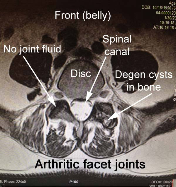
Below are examples of abnormal facet joints. In the image (below left), the lack of fluid in the facet along with the degenerative cysts can be seen.
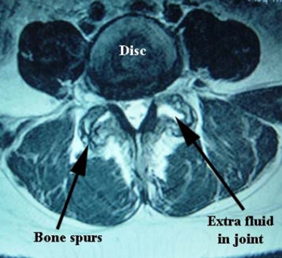
In this view (below right) of a spinal segment with facet degeneration, extra bone can be seen forming on one side (labeled “bone spurs”), and extra fluid is seen on the other since the two sides no longer fit well together. It is not hard to imagine that these joints don’t have smooth motion as the spine flexes and extends.
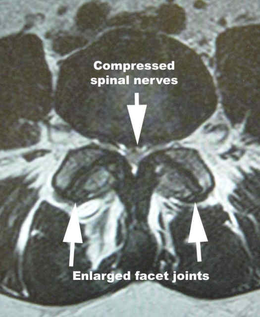
On this view, the facets also have no fluid signal in the joints, and they have overgrown to a point where they are compressing the nerves in the spinal canal.
This condition is called spinal stenosis.
On the side with arthritis, it's almost like the smooth joints have rusted.
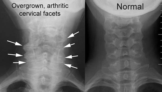
Cervical facet arthrosis
The facet joints in the back part of the cervical spine can also wear out in a similar manner.
In the images shown here which are AP views (looking from front to back), arthritic and enlarged facet joints can be compared to more normal appearing joints.
Treatment options
For cervical facet arthrosis, pain is generally treated non surgically. Options include the standard non operative treatments including medicine and physical therapy, but also sometimes:
Injections, and if they are successful,
Radiofrequency ablation (burning the nerves to the painful joints).